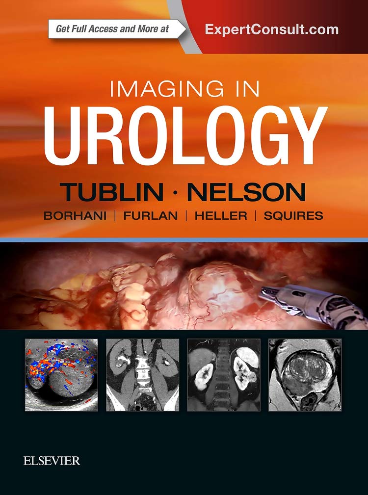
Imaging in Urology: 1ed
Written by a radiologist and a urologist, Imaging in Urology meets the needs of today’s urologists for a high-quality, highly relevant reference for evaluating and understanding the findings of radiologic exams related to urological disorders seen in daily practice. This unique title by Drs. Mitchell Tublin and Joel B. Nelson emphasizes the central role that imaging plays in the successful practice of urology by providing an image-rich review of urologic conditions ideal for both trainees and established urologists. Coverage includes introductory topics, imaging anatomy, and diagnoses, and tumor staging, all highlighted by about 1,600 images, drawings, and gross and microscopic pathology photos
- Focuses on imaging interpretation of the diagnostic entities that today’s urologist is likely to encounter in clinical practice
- Features a consistent, bulleted format highlighted by abundant images with detailed captions and annotations, all designed for quick reference at the point of care
- Covers key topics in urologic imaging, including the role of multiparametric MR in the staging and management of prostate carcinoma; the strengths and weaknesses of PI-RADS (Prostate Imaging Reporting and Data System); imaging approaches for characterization of the incidental adrenal lesion; and technical performance and utility of newer imaging modes such as CT urography, MR urography, and diffusion-weighted imaging
- Offers a focused, up-to-date method of meeting the AUA’s imaging expectations regarding imaging, which require training, review, and integration of ultrasound, CT, and other imaging modalities in the daily practice of urology
| Book | |
|---|---|
| Author | Tublin, Mitchell E. |
| Pages | 450 |
| Year | 2018 |
| ISBN | 9780323548090 |
| Publisher | Elsevier |
| Language | English |
| Uncategorized | |
| Edition | 1/e |
| Weight | 1.4 kg |
| Dimensions | 22.23 x 27.94 x 2.54 cm |
| Binding | Hardcover |


